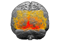
Brodmann area 18 is shown in orange in this image which also shows ares 17 (red) and 19 (yellow)
Human[]
Brodmann area 18, or BA18, is part of the occipital cortex in the human brain. It accounts for the bulk of the volume of the occipital lobe.
This area is also known as parastriate area 18. It is a subdivision of the cytoarchitecturally defined occipital region of cerebral cortex. In the human it is located in parts of the cuneus, the lingual gyrus and the lateral occipital gyrus (H) of the occipital lobe. Cytoarchitecturally it is bounded on one side by the striate area 17, from which it is distinguished by absence of a band of Gennari, and on the other by the peristriate area 19 (Brodmann-1909).
Guenon[]
Brodmann area 18 is a subdivision of the cerebral cortex of the guenon defined on the basis of cytoarchitecture. It is topographically and cytoarchitecturally homologous to parastriate area 18 of the human (Brodmann-1909). Distinctive features (Brodmann-1905): a wide, dense internal granular cell layer (IV); a distinct sublayer 3b of closely packed large pyramidal cells positioned in the external pyramidal layer (III) directly above layer IV; an almost cell free, narrow internal pyramidal layer (V) with no larger ganglion cells; a likewise very narrow, dense multiform layer (VI) composed of small polymorphic cells that form a distinct boundary with the underlying subcortical white matter. Like area 17 of Brodmann-1905, area 18 is relatively thin; the three deep layers are thin relative to the three outer layers; distinct boundaries between layers; abundance of granule cells; narrow layer VI; and sharp boundary between cortex and subcortical white matter.
External links[]
- For Neuroanatomy of the parastriate area 18 visit BrainInfo
- For Neuroanatomy of Brodmann area 18 visit BrainInfo
See also[]
| This neuroscience article is a stub. You can help the Psychology Wiki by expanding it. |
| This page uses Creative Commons Licensed content from Wikipedia (view authors). |