| Dura mater | ||
|---|---|---|
| Diagrammatic transverse section of the medulla spinalis and its membranes. (At border, dura mater is black line, arachnoid is blue line, and pia mater is red line.) | ||
| Latin | ' | |
| Gray's | subject #193 872 | |
| System | ||
| MeSH | A08.186.566.395 | |
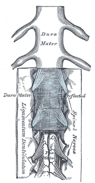
| The medulla spinalis and its membranes. | ||
The dura mater (from the Latin "hard mother"), or pachymeninx, is the tough and inflexible outermost of the three layers of the meninges surrounding the brain and spinal cord. The other two meningeal layers are the pia mater and the arachnoid mater. The dura mater isn't as tightly fitting around the spinal cord, extending past the spinal cord (which ends at L2, or second lumbar vertebra) to about the S2 (second sacral vertebra).
The dura mater has two layers: a deep layer and a superficial layer that is actually the skull’s inner periosteum, the membrane that lines bone. The dura separates into two layers at dural reflections, places where the inner dural layer is reflected as sheet-like protrusions into the cranial cavity.
There are two main dural reflections. The tentorium cerebelli exists between and separates the cerebellum and brainstem from the occipital lobes of the cerebrum (Shepherd, 2004). The falx cerebri, which separates the two hemispheres of the brain, is located in the longitudinal cerebral fissure between the hemispheres (Williams et al, 2002).
Channels called dural venous sinuses form between the two dural layers at dural reflections. These sinuses drain blood and cerebrospinal fluid from the brain and empty into the internal jugular vein. Meningeal veins, which course through the dura mater, and bridging veins, which drain the underlying neural tissue and puncture the dura mater, empty into these dural sinuses.
A subdural hematoma occurs when there is an abnormal collection of blood between the dura and the arachnoid, usually as a result of torn bridiging veins secondary to head trauma. An epidural hematoma is a collection of blood between the dura and the inner surface of the skull, and is usually due to arterial bleeding.
The American Red Cross and some other agencies accepting blood donations consider dura mater transplants, along with receipt of pituitary-derived growth hormone, a risk factor due to concerns about Creutzfeldt-Jakob disease.
References[]
- Shepherd S. 2004. "Head Trauma." Emedicine.com.
- Vinas FC and Pilitsis J. 2004. "Penetrating Head Trauma." Emedicine.com.
- Williams TH, Gluhbegovic N, Jew JY. 2002. "The Human Brain: Chapter 2: The Meninges and Blood Vessels of the Brain". University of Iowa.
- Red Cross: http://www.redcross.org/services/biomed/0,1082,0_553_,00.html
| Meninges of the brain and medulla spinalis |
|
Dura mater - Falx cerebri - Tentorium cerebelli - Falx cerebelli - Arachnoid mater - Subarachnoid space - Cistern - Cisterna magna - Median aperture - Cerebrospinal fluid - Arachnoid granulation - Pia mater |
de:Hirnhaut nl:Harde hersenvlies sv:dura mater
| This page uses Creative Commons Licensed content from Wikipedia (view authors). |
Assessment |
Biopsychology |
Comparative |
Cognitive |
Developmental |
Language |
Individual differences |
Personality |
Philosophy |
Social |
Methods |
Statistics |
Clinical |
Educational |
Industrial |
Professional items |
World psychology |
Biological: Behavioural genetics · Evolutionary psychology · Neuroanatomy · Neurochemistry · Neuroendocrinology · Neuroscience · Psychoneuroimmunology · Physiological Psychology · Psychopharmacology (Index, Outline)

