Assessment |
Biopsychology |
Comparative |
Cognitive |
Developmental |
Language |
Individual differences |
Personality |
Philosophy |
Social |
Methods |
Statistics |
Clinical |
Educational |
Industrial |
Professional items |
World psychology |
Biological: Behavioural genetics · Evolutionary psychology · Neuroanatomy · Neurochemistry · Neuroendocrinology · Neuroscience · Psychoneuroimmunology · Physiological Psychology · Psychopharmacology (Index, Outline)
| Ankle | ||
|---|---|---|
| Lateral view of the human ankle | ||
| Latin | articulatio talocruralis | |
| Gray's | subject #95 349 | |
| System | ||
| MeSH | A02.835.583.378.062 | |
| [[Image:|190px|center|]] | ||
- For a review of anatomical terms, see Anatomical position and Anatomical terms of location.
In human anatomy, the ankle joint is formed where the foot and the leg meet. The ankle, or talocrural joint, is a synovial hinge joint that connects the distal ends of the tibia and fibula in the lower limb with the proximal end of the talus bone in the foot. The articulation between the tibia and the talus bears more weight than between the smaller fibula and the talus.
Movement[]
The ankle joint is responsible for dorsiflexion (moving the toes up as when standing only on the heels) and plantar flexion of the foot (moving the toes down, as when standing on the toes), and allows for the greatest movement of all the joints in the foot. The ankle does not allow rotation.
In plantar flexion, the anterior ligaments of the joint become longer while the posterior ligaments become shorter. The reverse is true for dorsiflexion.
Articulation[]
The lateral malleolus of the fibula and the medial malleolus of the tibia along with the inferior surface of the distal tibia articulate with three facets of the talus. These surfaces are covered by cartilage.
The anterior talus is wider than the posterior talus. When the foot is dorsiflexed , the wider part of the superior talus moves into the articulating surfaces of the tibia and fibula, creating a more stable joint than when the foot is plantar flexed.
Ligaments[]
The ankle joint is bound by the strong deltoid ligament and three lateral ligaments: the anterior talofibular ligament, the posterior talofibular ligament, and the calcaneofibular ligament.
- The deltoid ligament supports the medial side of the joint, and is attached at the medial malleolus of the tibia and connect in four places to the sustentaculum tali of the calcaneus, calcaneonavicular ligament, the navicular tuberosity, and to the medial surface of the talus.
- The anterior and posterior talofibular ligaments support the lateral side of the joint from the lateral malleolus of the fibula to the dorsal and ventral ends of the talus.
- The calcaneofibular ligament is attached at the lateral malleolus and to the lateral surface of the calcaneus.
The joint is most stable in dorsiflexion and a sprained ankle is more likely to occur when the foot is plantar flexed. This type of injury more frequently occurs at the anterior talofibular ligament.
Name derivation[]
The word ankle or ancle is common, in various forms, to Germanic languages, probably connected in origin with the Latin "angulus", or Greek "αγκυλος", meaning bent.
Related terms[]
A common variant of the word "ankle" is "cankle", which is commonly used derogatorily to describe the ankles of obese individuals where the ankle and calf may be indistinguishable.
Fractures[]
Evaluation of ankle injuries for fracture is done with the Ottawa ankle rules, a set of rules that were developed to minimize unnecessary X-rays.
Additional images[]
")
")
")
")
")
References[]
- Calais-Germain, Blandine. "Anatomy of Movement", Eastland Press, 1993. ISBN 0-939616-17-3
- Martini, Frederic; Timmons, Michael; McKinnley, Michael. "Human Anatomy", 3rd Edition, Prentice-Hall, 2000. ISBN 0-13-010011-0
- Marieb, Elaine. "Essentials of Human Anatomy and Physiology", 6th Edition. Addison Wesley Longman, 2000. ISBN 0-8053-4940-5
See also[]
Look up this page on
Wiktionary:
Ankle
- Tarsus (skeleton)
External links[]
Joints and ligaments of lower limbs | |
|---|---|
| Coxal/hip |
iliofemoral - pubofemoral - ischiofemoral - head of femur - transverse acetabular |
| Knee-joint |
patellar - popliteal (oblique, arcuate) - collateral (medial/tibial, lateral/fibular) - cruciate (anterior, posterior) - menisci (medial, lateral) |
| Tibiofibular |
Superior tibiofibular: anterior of the head of the fibula - posterior of the head of the fibula |
| Talocrural/ankle |
deltoid - external lateral of the ankle-joint (anterior talofibular, posterior talofibular, calcaneofibular) |
| Foot - intertarsal |
Subtalar/talocalcaneal: anterior talocalcaneal - posterior talocalcaneal - lateral talocalcaneal - medial talocalcaneal - interosseous talocalcaneal |
| Foot - other |
Cuneonavicular, Cuboideonavicular, Intercuneiform and cuneocuboid, Tarsometatarsal/Lisfranc, Intermetatarsal, Metatarsophalangeal, Interphalangeal |
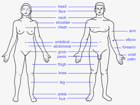 |
HEAD: Forehead – Eye – Ear – Nose – Mouth – Tongue – Teeth – Jaw – Face – Cheek – Chin TORSO: Shoulders – Spine – Chest – Breast – Ribcage – Abdomen – Belly button LIMBS: Arm – Elbow – Forearm – Wrist – Hand – Finger (Thumb - Index finger - Middle finger - Ring finger - Little finger) – Leg – Lap – Thigh – Knee – Calf – Heel – Ankle – Foot – Toe (Hallux) |
| This page uses Creative Commons Licensed content from Wikipedia (view authors). |