Assessment |
Biopsychology |
Comparative |
Cognitive |
Developmental |
Language |
Individual differences |
Personality |
Philosophy |
Social |
Methods |
Statistics |
Clinical |
Educational |
Industrial |
Professional items |
World psychology |
Biological: Behavioural genetics · Evolutionary psychology · Neuroanatomy · Neurochemistry · Neuroendocrinology · Neuroscience · Psychoneuroimmunology · Physiological Psychology · Psychopharmacology (Index, Outline)
| Brain: Pituitary stalk | ||
|---|---|---|
| Pituitary stalk not labeled, but is the vertical blue portion. | ||
| Basal view of a human brain (Infundibulum labeled third from the top on right) | ||
| Latin | infundibulum neurohypophyseos | |
| Gray's | subject #189 813 | |
| Part of | ||
| Components | ||
| Artery | ||
| Vein | ||
| BrainInfo/UW | hier-388 | |
| MeSH | A06.407.747 | |
- Also see infundibulum (disambiguation) for other uses of the term.
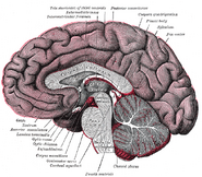
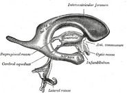
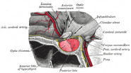
The pituitary stalk (also known as the infundibular stalk or simply the infundibulum) is the connection between the hypothalamus and the posterior pituitary. The floor of the third ventricle is prolonged downward as a funnel-shaped recess, the infundibular recess, into the infundibulum, and to the apex of the latter the hypophysis or pituitary is attached.[1]
It carries axons from the magnocellular neurosecretory cells of the hypothalamus down to the posterior pituitary where they release their neurohypophysial hormones, oxytocin and vasopressin, into the blood.
This connection is called the hypothalamo-hypophyseal tract or hypothalamo-neurohypophyseal tract.
Compression[]
It has been thought that the pituitary stalk may become compressed due to suprasellar tumors in the pars tuberalis region, and that the resulting compression may cause hyperprolactinemia.[2] This phenomenon has been described as the stalk effect or pituitary stalk compression syndrome.[2]
However, at least one article suggests that the increase in prolactin in these cases may instead be caused by the tumor's secretion of preprotachykinin A derived tachykinins, substance P, and/or neurokinin A.[2]
Additional images[]
External links[]
References[]
| This page uses Creative Commons Licensed content from Wikipedia (view authors). |


