Assessment |
Biopsychology |
Comparative |
Cognitive |
Developmental |
Language |
Individual differences |
Personality |
Philosophy |
Social |
Methods |
Statistics |
Clinical |
Educational |
Industrial |
Professional items |
World psychology |
Biological: Behavioural genetics · Evolutionary psychology · Neuroanatomy · Neurochemistry · Neuroendocrinology · Neuroscience · Psychoneuroimmunology · Physiological Psychology · Psychopharmacology (Index, Outline)
| Brain: Brodmann area 25 | ||
|---|---|---|
| Brodmann area 25 is shown in orange. | ||
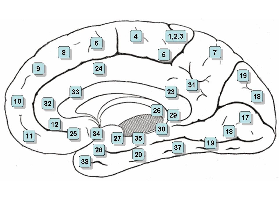 | ||
| Medial surface of the brain with Brodmann's areas numbered. | ||
| Latin | Area subgenualis | |
| Gray's | subject # | |
| Part of | ||
| Components | ||
| Artery | ||
| Vein | ||
| BrainInfo/UW | - | |
| MeSH | [1] | |
Sagittal MRI slice with highlighting indicating location of the subgenual anterior cingulate cortex.
Brodmann area 25 (BA25) is an area in the cerebral cortex of the brain and delineated based on its cytoarchitectonic characteristics. It is also called the subgenual area, area subgenualis or subgenual cingulate. It is the 25th "Brodmann area" defined by Korbinian Brodmann (thus its name). BA25 is located in the cingulate region as a narrow band in the caudal portion of the subcallosal area adjacent to the paraterminal gyrus. The posterior parolfactory sulcus separates the paraterminal gyrus from BA25. Rostrally it is bound by the prefrontal area 11 of Brodmann.[1]
History[]
Brodmann described this area as it is labeled now in 1909. Originally in 1905, Brodmann labeled the area as part of area 24. In 1909, he divided the area into area 24 and 25.[2]
Function[]
This region is extremely rich in serotonin transporters and is considered as a governor for a vast network involving areas like hypothalamus and brain stem, which influences changes in appetite and sleep; the amygdala and insula, which affect the mood and anxiety; the hippocampus, which plays an important role in memory formation; and some parts of the frontal cortex responsible for self-esteem.[3]
Pathology[]
One study has noted that BA25 is metabolically overactive in treatment-resistant depression and has found that chronic deep brain stimulation in the white matter adjacent to the area is a successful treatment for some patients.[4] A different study found that metabolic hyperactivity in this area is associated with poor therapeutic response of persons with Major Depressive Disorder to cognitive-behavioral therapy and venlafaxine.[5]
Image[]
Notes and references[]
- ↑ subgenual area 25. braininfo.rprc.washington.edu, retrieved November 18, 2006.
- ↑ area 25 of Brodmann-1909. braininfo.rprc.washington.edu, retrieved November 19, 2006.
- ↑ "Faulty Circuits", Scientific American, April 2010
- ↑ Deep Brain Stimulation for Treatment-Resistant Depression neuron.org, March 3, 2005. Retrieved November 18, 2006.
- ↑ Predictors of nonresponse to cognitive behavioural therapy or venlafaxine using glucose metabolism in major depressive disorder cma.ca, May 2009. Retrieved May 23, 2009.
See also[]
| This page uses Creative Commons Licensed content from Wikipedia (view authors). |