Assessment |
Biopsychology |
Comparative |
Cognitive |
Developmental |
Language |
Individual differences |
Personality |
Philosophy |
Social |
Methods |
Statistics |
Clinical |
Educational |
Industrial |
Professional items |
World psychology |
Biological: Behavioural genetics · Evolutionary psychology · Neuroanatomy · Neurochemistry · Neuroendocrinology · Neuroscience · Psychoneuroimmunology · Physiological Psychology · Psychopharmacology (Index, Outline)
The telencephalon (te-len-seff-a-lon) is the technical name for a large region within the brain which is attributed many functions, which some groups would class as unique features which make humans stand out from other species. Many people refer to it as the cerebrum, but due to naming conventions of organs, is technically referred to as the telencephalon.
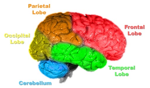
The lobes of the cerebral cortex include the frontal (red), temporal (green), occipital (yellow), and parietal lobes (orange). The cerebellum (blue) is not part of the telencephalon.
As a more technical definition, the telencephalon refers to the cerebral hemispheres and other, smaller structures within the brain, despite the fact that the telencephalon is one of the larger divisions (in terms of number). This term is technically the anterior-most embryological division of the brain that develops from the prosencephalon.
Structure[]
The telencephalon, in name, refers to the region of the brain which is composed of the following sub-regions;
- Limbic System
- Cerebral Cortex or Cortices of the cerebral hemispheres.
- Basal Ganglia
- Corpus Striatum
- Olfactory Bulb
Composition[]
The telencephalon comprises what most people think of as the "brain". It lies on top of the brainstem and is the largest and most well-developed of the 5 major divisions of the brain. Phylogenetically it is the newest structure, with mammals having the largest and most well-developed among all species. It emerges from the prosencephalon, the first of three vesicles that form from the embryonic neural tube.
Originally,indeed there was thought to be four divisions of the telencephalon, although through later reasearch other sub-divisions were described. This four-division scheme is referred to as the traditional division
In humans, the telencephalon surrounds older parts of the brain. Limbic, olfactory, and motor systems project fibers from subcortical (deeper) areas of the cerebrum to parts of the brainstem. Cognitive and volitive systems project fibers from cortical areas of the cerebrum to thalamus and to other regions of the brainstem. The neural networks of the telencephalon facilitate complex learned behaviors, such as language.
The cerebrum is the largest part of the brain, and contains white matter and grey matter. Grey matter is highly folded; functionally this is thought to allow a greater number of cells in the same volume due to the increase in its surface area. The telencephalon includes regions of archipalliar, paleopalliar, and neopalliar origin. Profound development of the neopallium, which comprises the cerebral cortex, is unique among humans and Old World monkeys.
Functions[]
Note: As the telencephalon is a gross division with many subdivisions and sub-reigons, it is important to state that this section lists the functions which the telencephalon as a whole serves.
Language and communication[]
Speech and language are mainly attributed to parts of the cerebral cortex, which is one portion of the telencephalon. Motor portions of language are attributed to Broca's area within the frontal lobe. Speech comprehension is attributed to Wernicke's area which lies at the temporal-parietal lobe junction. These two regions are interconnected by a large white matter tract known as the arcuate fasciculus.
Movement[]
The telencephalon is the part of the brain which attributes motor function to the body. These functions originate within the primary motor cortex and other frontal lobe motor area. In many cases when this part of the brain is damaged the brain has an inability to send signals to nerves that innervate muscles motoneurons, and can lead to diseases such as Motor Neurone Disease. This kind of damage results in loss of muscular power and precision rather than total paralysis. This is because there are other, older portions of the brain that also subserve motor function.
Olfaction[]
The olfactory bulb lies on the underside of the most anterior portion of the brain. This is a rather large portion of the telencephalon in most mammals. However in humans this part of the brain has become orders of magnitude smaller as other functions have taken over after being proven more evolutionarily advantageous. Damage to this portion of the brain results in a loss of the sense of smell.
Memory[]
Memory formation is associated with the hippocampus. This association was originally described after a patient (HM) had both his hippocampuses (left and right) surgically removed to treat severe epilepsy. After surgery this patient suffered from anterograde amnesia, or the inability to form new memories. This problem is also addressed slightly in the film Memento, in which the protagonist has to take pictures of people he has met in order to be able to remember what to do in the days following his accident, so in that respect, the film is factually accurate.
Emotion[]
Emotional functions are attributed to a wide network of telencephalic and other regions grouped together as the limbic system. The amygdala is a nucleus known to contribute a great deal to the emotion of fear. This region is part of the Papez circuit, which is the anatomical loop between various brain regions responsible for cortical control of information.
Programmed cell death[]
Purpose[]
Programmed Cell Death (PCD) is not uncommon within the telencephalon or it's sub-regions. It is thought to be one of the processes by which growth and differentation grows, and is a universal feature of the embryonic and postnatal central nervous system [1], and has been noted to be at work within the telencephalons of animals such as rats, mice, and other vermin. In some animals such as the monkey, over 50% of neurons within their cerebral cortex have been found to be affected by PCD during early stages of life. This is thought to solicit growth of the brain due to the grown of the size of the cranium and other parts of the body expected to grow throughout the life cycle of a monkey.
The main reason for PCD is because cells need to die so other ones can be made, for spatial reasons. If a neuron does not establish the correct synaptic connection, it will die shortly thereafter. This is seen as some form of "competition" within the space of the telencephalon and is a form of the "survival of the fittest". However there are exceptions to the rule; within rats some cells are even programmed die during proliferation within the ventricular zones of the telencephalon. It is thought that this is at a stage during which axons are not yet formed nor synaptically connected.
Effects[]
PCD within the brain affects glial cells and neurons through apoptosis (4). According to extensive research on rodents this period is usually during developmental or adolescent stages. During this time the regeneration process can take place because the "materials" and environment are a perfect breeding ground for cell regeneration.
Stages[]
During the stages of apoptosis, which seems to constitute the majority of PCD within the brain, various morphological changes occur, such as:
- Cell shrinkage.
- Membrane blebbage, or inconsistency within the structure of the membrane.
- Pyknosis, or a condensation of chromatin within the nucleus.
- Karyorrhexis, or fragmentation of the nuclei.
- Organelles and plasma membrane remain intact throughout the process.
- A lack of inflammation.
- Removal by microphages or adjacent glial cells.
Of course, there are some differences to these stages, but they are relatively similar in practice. The apoptosis within the telencephalon can also be characterised distinctly by it's DNA pattern, which becomes fragmented into oligonucleosomal fragments of around 180-200 pairs. These give a typical "ladder" pattern when viewed on or in agarose gel electrophoresis.
Cell regeneration[]
Xenopus laevis[]
Larval stage[]
In a study of the telencephalon conducted in Hokkaido University on African clawed frogs (xenopus laevis)[2], it was discovered that during larval stages the telencephalon was able to regenerate around half of the anterior portion (otherwise known as partially truncated), after a reconstruction of a would-be accident, or malformation of features.
The actual regeneration and active proliferation of cells within the clawed frog is quite remarkable; regenerated cells being almost functionally identical to the ones originally found in the brain after birth despite the obvious lack of brain matter for a sustained period of time.
This kind of regeneration is completely dependant upon ependymal layer cells covering the cerebral lateral ventricles, within a short period before, or within the initial stage of wound healing. This is observed within the stages of healing within larvae of the clawed frog.
Developed stage[]
Unfortunately the regeneration within the developed stage of the clawed frog is completely different than within the larval stage. Because the cells adhere to one another they are unable to form an entity which is able to cover the cerebral lateral ventricles. Thus the telencephalon remains truncated and the loss of function becomes permanent.
Effects of abnormality[]
After removing over half of the telencephalon in the developed stage of the clawed frog, the lack of functions within the animal was apparent, manifesting with obvious difficulties in movement, nonverbal communication between other species, as well as other difficulties thought to be similar to those seen in humans.
This kind of regeneration is still relatively unknown in regard to regeneration within larval stages, similar to the human fetal stage.
References[]
- Template:Anb Yoshino J, Tochinai S. Successful reconstitution of the non-regenerating adult telencephalon by cell transplantation in Xenopus laevis. Dev Growth Differ. 2004;46(6):523–34. PMID 15610142
- Template:Anb Levi-Montalcini, R. (1949) Proliferation, differentiation and degeneration in the spinal ganglia of the chick embryo under normal and experimental conditions. Pages 450 - 502
- Template:Anb Yaginuma, H., Tomita, M., Takashita, N., McKay, S., Cardwell, C., Yin, Q.- Aminobuytric acid immunoreactivity within the human cerebral cortex. Pages 481 - 500
- Template:Anb Haydar, T. F, Kuan, C., Y., Flavell, R. A. & Rakic, P. (1999) The role of cell death in regulating the size and shape of the mammalian forebrain. Pages 621 - 626
See also[]
| This page uses Creative Commons Licensed content from Wikipedia (view authors). |