Assessment |
Biopsychology |
Comparative |
Cognitive |
Developmental |
Language |
Individual differences |
Personality |
Philosophy |
Social |
Methods |
Statistics |
Clinical |
Educational |
Industrial |
Professional items |
World psychology |
Biological: Behavioural genetics · Evolutionary psychology · Neuroanatomy · Neurochemistry · Neuroendocrinology · Neuroscience · Psychoneuroimmunology · Physiological Psychology · Psychopharmacology (Index, Outline)
| Utricle (ear) | ||
|---|---|---|
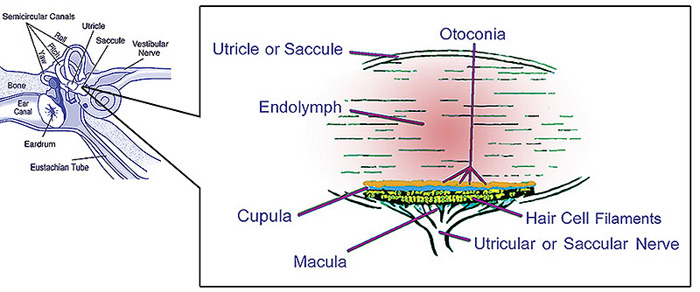 | ||
| illustration of otolith organs showing detail of utricle, otoconia, endolymph, cupula, macula, hair cell filaments, and saccular nerve | ||
| Latin | utriculus | |
| Gray's | subject #232 1051 | |
| System | ||
| MeSH | A09.246.631.909.625 | |
| [[Image:{{{Image2}}}|190px|center|]] | ||
| {{{Caption2}}} | ||
The utricle, or utriculus, along with the saccule is one of the two otolith organs located in the vertebrate inner ear.
Anatomy[]
The utricle is larger than the saccule and is of an oblong form, compressed transversely, and occupies the upper and back part of the vestibule, lying in contact with the recessus ellipticus and the part below it.
The utricle contains mechanoreceptors called hair cells that distinguish between degrees of tilting of the head, thanks to their apical cilia set-up. These are covered by otolith and, once you tilt your head, otolith viscosity has the cilia tilt as well. Depending on whether the tilt is in the direction of the kinocilium or not, the resulting hair cell polarisation is excitatory (depolarising) or inhibitory (hyperpolarisation), respectively. This signal to the vestibular nerve (which takes it to the brainstem) does not adapt with time, so if you're lying in bed, you still feel as if you're lying in bed 9 hours afterwards when you wake up.
That portion which is lodged in the recess forms a sort of pouch or cul-de-sac, the floor and anterior wall of which are thickened, and form the macula acustica utriculi, which receives the utricular filaments of the acoustic nerve.
The cavity of the utricle communicates behind with the semicircular ducts by five orifices.
From its anterior wall is given off the ductus utriculosaccularis, which opens into the ductus endolymphaticus.
Additional images[]
")
")
See also[]
External links[]
This article was originally based on an entry from a public domain edition of Gray's Anatomy. As such, some of the information contained herein may be outdated. Please edit the article if this is the case, and feel free to remove this notice when it is no longer relevant.
Sensory system: Auditory and Vestibular systems (TA A15.3, GA 10.1029) | |||||||||||||||
|---|---|---|---|---|---|---|---|---|---|---|---|---|---|---|---|
| Outer ear |
Pinna (Helix, Antihelix, Tragus, Antitragus, Incisura anterior auris, Earlobe) • Ear canal • Auricular muscles | ||||||||||||||
| Middle ear |
| ||||||||||||||
| Inner ear/ (membranous labyrinth, bony labyrinth) |
| ||||||||||||||
| {| class="navbox collapsible nowraplinks" style="margin:auto; " | |||||||||||||||
| |||||||||||||||
|}
| This page uses Creative Commons Licensed content from Wikipedia (view authors). |