Assessment |
Biopsychology |
Comparative |
Cognitive |
Developmental |
Language |
Individual differences |
Personality |
Philosophy |
Social |
Methods |
Statistics |
Clinical |
Educational |
Industrial |
Professional items |
World psychology |
Biological: Behavioural genetics · Evolutionary psychology · Neuroanatomy · Neurochemistry · Neuroendocrinology · Neuroscience · Psychoneuroimmunology · Physiological Psychology · Psychopharmacology (Index, Outline)
| Zonule of Zinn | ||
|---|---|---|
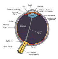
| Schematic diagram of the human eye. (Zonular fibers labeled at upper left.) | ||
| Latin | zonula ciliaris | |
| Gray's | subject #226 1018 | |
| System | ||
| MeSH | [1] | |
| The upper half of a sagittal section through the front of the eyeball. (Zonule of Zinn visible near center.) | ||
The zonule of Zinn (Zinn's membrane, ciliary zonule) is a ring of fibrous strands connecting the ciliary body with the crystalline lens of the eye. The zonule is split into two layers: a thin layer which lines the hyaloid fossa and a thicker layer which is a collection of zonular fibers. Collectively, the fibers are known as the suspensory ligament of the lens[1].
Overview[]
The zonular fibers pass over the ciliary body and are attached to the capsule of the lens a short distance in front of its equator. These fibers change the focusing power of the eye by changing the tension of the fibers by contraction and relaxation of the ciliary muscle.
It should not be confused with the Annulus of Zinn, though it is named after the same person (Johann Gottfried Zinn).
The zonule is essentially a system of numerous fibers that run from the ciliary body to the lens periphery, whose function is both securing the lens in the optical axis and transferring forces from the ciliary muscle in accommodation. Its exact anatomy and morphology is yet to be fully understood, due mainly to the fact that it is very difficult to study both in-vivo and ex-vivo. In an in-vivo setting, the ciliary zonule is some 3 mm behind the cornea, with the iris shielding it from any direct optical examination. Furthermore, zonular fiber dimensions are in the order of tens of micrometres, requiring high magnification instruments. Recent in-vivo studies have relied mostly on ultrasound biomicroscopy (Ludwig, Wegscheider et al. 1999), but they suffer from insufficient resolution and movement artifact. For this reason, ex-vivo investigation is currently the only option for the study of the zonule to date.
Initial anatomical studies of the zonule were made in vivo with a slit lamp in patients with colobomata (holes) of the iris (McCulloch 1954). Descriptions varied depending on the source, but in general, the interpretation obtained was that of a broad belt surrounding the lens equator, with its anterior surface running from the lens to the ciliary processes and its back face forming the posterior boundary of the posterior chamber. Wislocki (Wislocki 1952) incorporated histological preparation with fixing agents and/or stains like aniline blue, periodic acid-Schiff and Flemming’s solution to the study of the zonule. This allowed for the fibrous characteristic of the zonule to be seen.
The study of the zonular architecture then experienced a leap with the advent and application of the scanning electron microscope (SEM) and transmission electron microscope (TEM). SEM and TEM allowed great magnification (100,000+), high resolution and high contrast images of tissue. TEM studies demonstrated that the zonular strands were not artifacts but were really micrometre sized fibers made of microfibrils (Streeten 1982), but it was SEM that proved to be an invaluable tool for describing the three dimensional architecture of the zonule due to its high depth of field. The first SEM observations in this field came from Hansson (Hansson 1970) in rat eyes, followed by a great effort in the seventies and eighties by various scientists to describe the spatial arrangement of the zonule in humans and primates (Raviola 1971; Bornfeld, Spitznas et al. 1974; Davanger 1975; Erckenbrecht and Rohen 1975; Farnsworth, Mauriello et al. 1976; Streeten 1977; Rohen 1979; Streeten 1982). Their observations are, in general, what is accepted to be the zonular architecture, and will be described in detail next.
The components of the suspensory apparatus of the lens run a complex but continuous course from the ora serrata to the edge of the lens, and are divided in the literature into four major sections: The pars orbicularis, which lies in the pars plana; the zonular plexus between the ciliary processes; the zonular fork which is the branching of the fibers at the mid zone of the ciliary valleys, and the limbs (anterior, posterior and equatorial) of the zonule (Bron, Tripathi et al. 1997). The last section is of particular importance to the architecture of the accommodation apparatus, given that it describes the anchoring points and routes of the different zonular fibers from the ciliary body to the lens.
The majority of the zonules have origin in the posterior end of the pars plana. They run forward like a mat until they reach the pars plicata, where they divide into different zonular plexuses between the valleys of the ciliary processes, attaching closely to their walls with secondary tension fibers acting as a fulcrum (pivot point). Continuing anteriorly on the pars plicata, each plexus divides into a fork, consisting of three fiber groups running to the anterior, posterior and equatorial lens capsule (Rohen 1979). The anterior zonule is described as running mainly from the pars plana to the anterior periphery of the lens, with some supporting fibers that originate in the pars plicata. The posterior zonule runs predominantly from the pars plicata to the post equatorial lens with supporting fibers emerging from the pars plana. The equatorial zonule passes from the pars plicata to the lens equator (Bornfeld, Spitznas et al. 1974). The pre-equatorial (anterior), equatorial and post-equatorial insertions of the zonular fibers into the lens capsule are different. The pre-equatorial insertion of the anterior zonules is relatively dense, as they all insert in approximately the same area 1.5 mm away from the equator in 25-60 µm wide bundles. When merging with the capsule, the zonular insertion flattens, and splits into smaller strands that fan out and insert into the capsule forming the zonular lamella, which in average continues 0.5 mm central to the insertion site (Streeten 1977). The space between the anterior and posterior zonule is called the canal of Hannover, and is populated by equatorial and meridional fibers which are fewer and thinner than the anterior zonules. Equatorial fibers form bundles of 10-15 µm, which also fan out and insert into the lens capsule, causing striations along the lens capsule (Streeten 1977). The posterior fibers insert in various layers over a 0.4 to 0.5 mm wide zone. Anteriorly, they insert at the posterior edge of the lens equator, and posteriorly they can extend up to 1.25 mm from the equatorial margin (Bron, Tripathi et al. 1997). The posterior zonules appear less organized and developed than the anterior ones, but this is attributed to the fact that they insert at various levels, with the hyaloid membrane being the posterior limit.
When colour granules are displaced from the Zonules of Zinn (by friction against the lens), the irises slowly fade. In some cases those colour granules clog the channels and lead to Glaucoma Pigmentosa.
The zonules are primary made of fibrillin, a connective tissue protein.[2] Mutations in the fibrillin gene lead to the condition Marfans Syndrome, and consequences include an increased risk of lens dislocation.[2]
Clinical appearance[]
The zonules of Zinn are difficult to visualize using a slit lamp, but may be seen with exceptional dilation of the pupil, or if a coloboma of the iris or a suluxation of the lens is present.[3] The number of zonules present in a person appears to decrease with age.[4] The zonules insert around the outer margin of the lens (equator), both anteriorly and posteriorly.[5]
Additional Images[]
References[]
- ↑ Vision - via the optic nerve (CN II)
- ↑ 2.0 2.1 Cite error: Invalid
<ref>tag; no text was provided for refs namedAdlersPhysiology - ↑ McCulloch, C (1954-1955). The Zonule of Zinn: its Origin, Course, and Insertion, and its Relation to Neighboring Structures.. Transactions of the American Ophthalmological Society 52: 525–85.
- ↑ Cite error: Invalid
<ref>tag; no text was provided for refs namedScannningEM - ↑ Farnsworth, PN, Mauriello, JA; Burke-Gadomski, P; Kulyk, T; Cinotti, AA (1976 Jan). Surface ultrastructure of the human lens capsule and zonular attachments.. Investigative ophthalmology 15 (1): 36–40.
External links[]
- Diagram at unmc.edu
- Diagram at eye-surgery-uk.com
- Diagram and overview at webschoolsolutions.com
- Dictionary at eMedicine ciliary+zonule
- Histology at Boston University 08011loa
This article was originally based on an entry from a public domain edition of Gray's Anatomy. As such, some of the information contained herein may be outdated. Please edit the article if this is the case, and feel free to remove this notice when it is no longer relevant.
| Sensory system - Visual system - Eye - edit |
|---|
| Anterior chamber | Aqueous humour | Blind spot | Choroid | Ciliary body | Conjunctiva | Cornea | Iris | Lens | Macula | Optic disc | Optic fovea | Posterior chamber | Pupil | Retina | Schlemm's canal | Sclera | Tapetum lucidum | Trabecular meshwork | Vitreous humour |
| This page uses Creative Commons Licensed content from Wikipedia (view authors). |
